
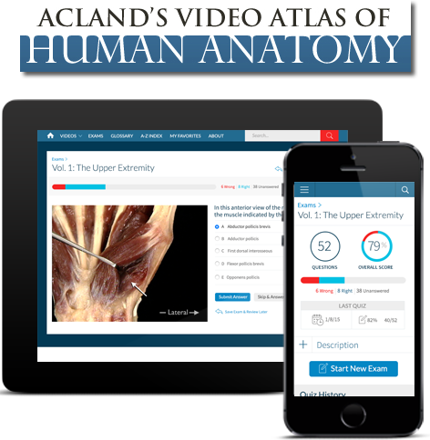
Acland’s Video Atlas of Human Anatomy
Acland’s Video Atlas of Human Anatomy is the optimal anatomical learning solution. Watch and listen to world renowned anatomist Dr. Robert Acland — Professor Emeritus of Surgery, University of Louisville School of Medicine — as he shows and explains anatomical movement within each region of the body covering bones, muscles, tendons, organs, and more.
Available online, this atlas is a perfect resource for any institution’s residents, faculty and students within medical, nursing, physician assistant, physical/occupational therapy, speech/language, and dentistry programs. Acland’s Video Atlas of Human Anatomy provides foundational information to supplement the learning and teaching they receive in the classroom and in practice.
Real Movement
Exquisite Dissections
Acland's Video Atlas of Human Anatomy contains nearly 330 videos of real human anatomic specimens in their natural colors, including 5 new, groundbreaking videos of the inner ear.
Dr. Robert Acland presents moving structures - muscles, tendons, and joints - making the same movements that they make in life. The videos show complex structures step by step - from bone to surface anatomy - to provide a foundation for understanding anatomical structure and function. The entire series was digitally re-mastered producing clearer, brighter, and more detailed videos than seen in previous versions.
Unique Features
Fresh human specimens in their natural colors
The Video Atlas images are direct video recordings of real human anatomic specimens.

Exquisite dissections
The dissections were done by skilled clinical anatomists, using the finest surgical techniques.
Clear narration and labeled structures
A concise narration runs throughout the program, using the simplest possible language.
Building complex structures step-by-step
In each part of the body, the Video Atlas starts with structures that give you the foundation for your understanding.
Why Acland’s Video Atlas of Human Anatomy?
Supports the teaching and learning of anatomy with more than 300 narrated videos
Includes upper extremity, lower extremity, trunk, head & neck, and internal organs; plus, brand new groundbreaking inner ear content
Demonstrates muscle function in 3D to colleagues, staff, and patients
Enables downloading of PDF transcripts for use in teaching guides and patient handouts
Ability to browse video clips, share with colleagues, and add to a “favorites” area for quick reference
Allows students independent, frequent viewing for review, self-assessment, and practical exam preparation — as well as tracking their performance using interactive Q&A and timed or review quizzes
5-Volume Gross Anatomy Videos
These gross anatomy videos are comprised of 5 volumes, organized by region. Each volume offers in-depth coverage of the bones, joints, muscles, and more. This online resource also offers material to support student and faculty learning and teaching structure identification including self-assessment/Q&A, PDF transcripts of the videos used for handouts, and more.
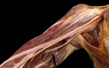
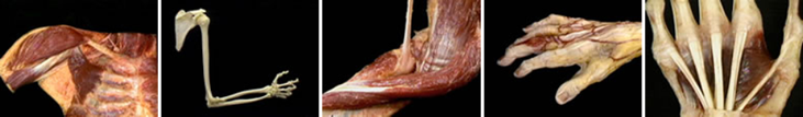
The Upper Extremity
Volume 1 has three sections: 1) the shoulder, 2) the arm and forearm, and 3) the hand. Each section shows first the bones and the movements they can make, then the joints and the ligaments that limit their movements, and then the muscles and the movements they produce. Once these major structures are understood, the blood vessels and nerves are added to the picture.
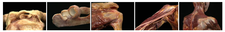
The section on the shoulder includes structures that are essential to the upper extremity, but that are often taught as parts of either the trunk or the neck. These include the muscles that move the scapula, the brachial plexus, and the subclavian blood vessels.
Volume 1
The Upper Extremity
Please select the video you want to watch:
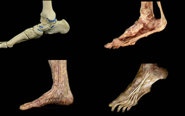
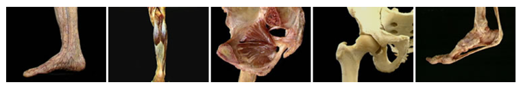
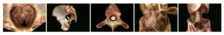
The Lower Extremity
Volume 2 has four sections: 1) the hip, 2) the knee, 3) the ankle, and 4) the foot. Each section shows first the bones and the movements they can make, then the joints and the ligaments that limit their movements, and then the muscles and the movements they produce. Once these major structures are understood, the blood vessels and nerves are added to the picture.
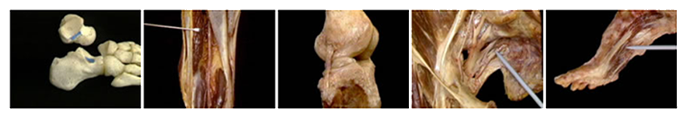
The section on the hip includes a full account of the bony pelvis. The first three sections are focused on a joint and the muscles that move it. Muscles that produce movements at two joints are shown in both sections. The effect of any muscle action is shown both when the extremity is bearing weight and when it is free to move.
Volume 2
The Lower Extremity
Please select the video you want to watch:
The Trunk (Musculoskeletal System)
Volume 3 has four sections: 1) the spine, 2) the thorax, 3) the abdomen, and 4) the pelvis.
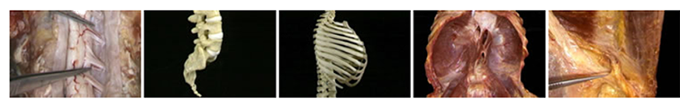
Section 1 shows the vertebral column, the paraspinous muscles, and the spinal cord. Section 2 shows the thorax as the structure that contains the heart and lungs, including the dynamic anatomy of respiratory movement. Sections 3 and 4 show the musculoskeletal anatomy of the abdomen and pelvis, as the upper and lower parts of the "container" for the abdominal and pelvic viscera. Sections 2, 3, and 4 also show the major blood vessels and nerves of their respective regions.
Volume 3
The Trunk
Please select the video you want to watch:
The Head and Neck
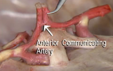
Volume 4 has 11 sections: 1) support and movement of the head, 2) the skull and facial skeleton, 3) the nasal cavity and associated structures, 4) the oral cavity and associated structures, 5) the larynx and associated structures, 6) the facial muscles and the scalp, 7) the brain and its surroundings, 8) the nerves of the head and neck, 9) the blood vessels of the head and neck, 10) the eye and its surroundings, and 11) the ear.
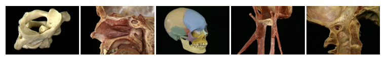
Section 1 shows the cervical spine and the musculoskeletal structures that connect the head to the body. Section 2 gives a highly three-dimensional display of the challenging bony anatomy of the skull and facial skeleton. Sections 3, 4, and 5 focus on the major "visceral" parts of the head and neck that are associated with breathing, eating, and speaking.
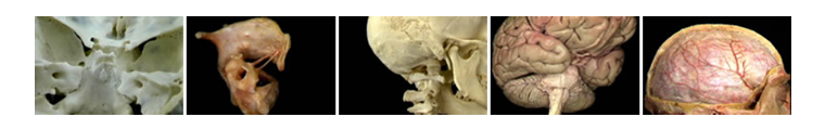
Section 6 shows the muscles involved in facial movement. Section 7 covers the external features of the brain and its relation to the cranial cavity. Section 8 shows the intracranial and extracranial course of the twelve cranial nerves. Section 9 shows both the intracranial and extracranial arteries and veins, including the venous sinuses. Section 10 shows the structures of the orbital cavity, the eyelids, and the external features of the eye. Section 11 shows the external and middle ear, including the dynamic anatomy of the ear drum and auditory ossicles.
AccessVolume 4
The Head and Neck
Please select the video you want to watch:
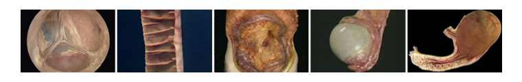
The Internal Organs and Reproductive System
Volume 5 has three sections: 1) the thoracic organs, 2 the abdominal organs, and 3) the reproductive system.
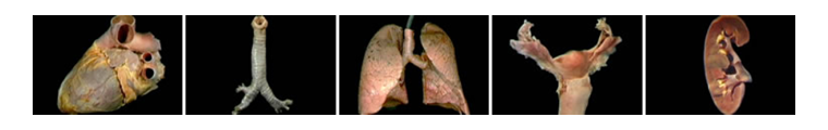
Section 1 includes a highly three-dimensional display of the heart, including internal views of the four chambers of the heart, and action shots of the valves in motion. Section 2 shows the stomach and intestines (with developmental animations explaining the rotation of the midgut), the liver and pancreas, and the kidneys and urinary system. Section 3 shows the internal and external features of the male and female reproductive organs.
Volume 5
The Internal Organs
Please select the video you want to watch:

Contact
Contact Ovid Technical Support
Our Technical Support and Customer Service Specialists are here to help. Please fill the following form to get in touch with us!